
Medical Image Gallery
Ortho Gallery
Anatomical illustrations, clinical X-Rays, MRI scans, and patient recovery photography.
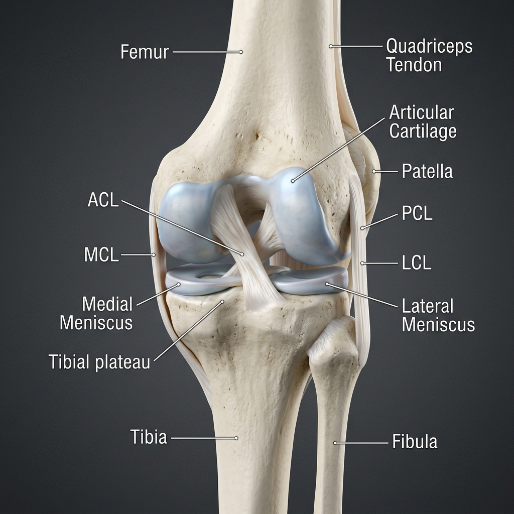 Anatomy
Anterior Knee Anatomy & Joint Capsule
Detailed 3D view of patella, meniscus, cartilage surfaces and ligament attachments.
Added: 2026-08-10
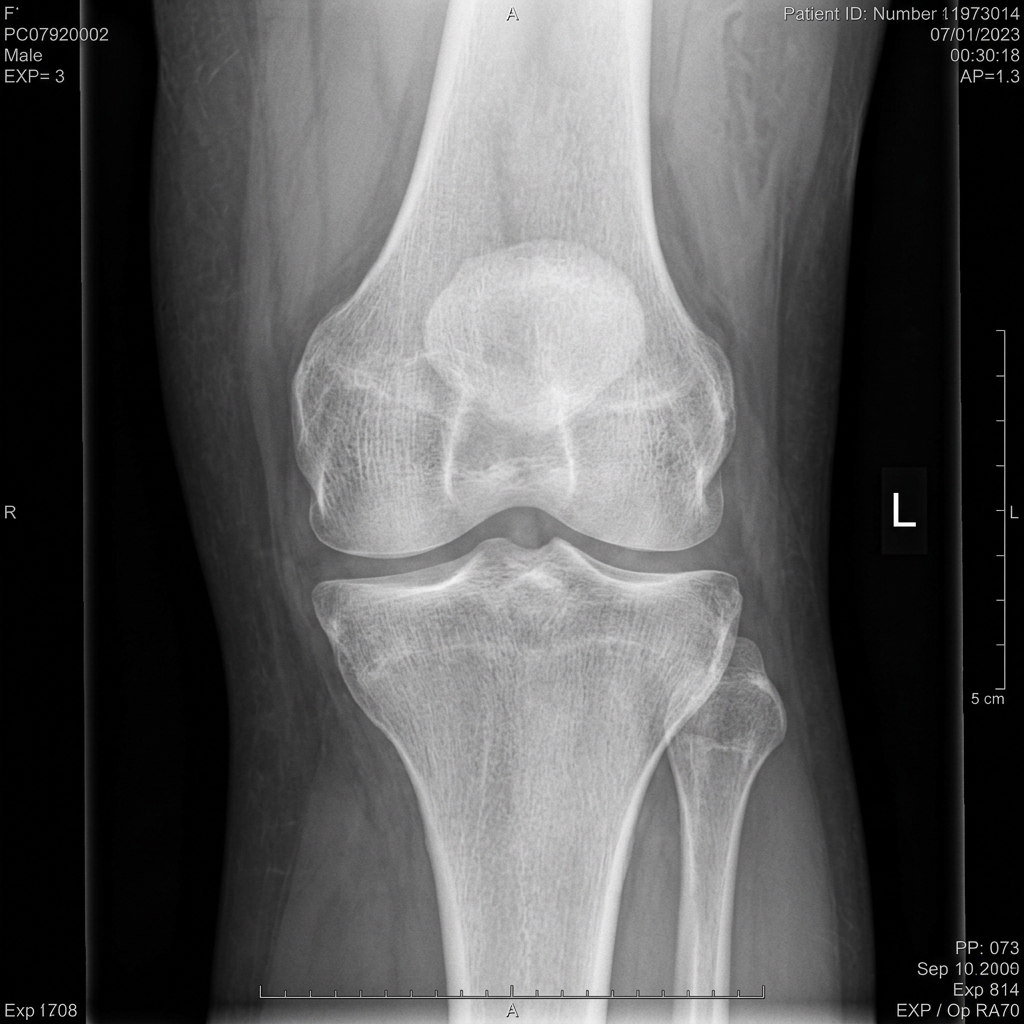 X-Ray
X-RayX-Ray Assessment: Patellofemoral Alignment
Radiograph showing patellar alignment and joint space width.
Added: 2026-08-08
MRIMRI Scan: Knee Joint Cartilage & Meniscus
Sagittal view showing medial meniscus posterior horn and femoral cartilage.
Added: 2026-08-05
X-RayLumbar Spine Vertebral Alignment
Lateral spine radiograph depicting L1-L5 lordotic curvature and disc heights.
Added: 2026-08-01